Research
Develpopment of a continuous egdeless crystal pet system for breast cancer
Develpopment of a continuous egdeless crystal pet system for breast cancer
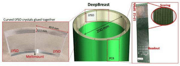
In this project we propose to build a novel continuous-scintillator Positron Emission Tomography (PET) scanner devoted to breast cancer diagnostic and therapy follow-up. This new concept of PET is designed to perform better than the current PET scanners in terms of sensitivity, spatial resolution, and uniformity throughout its field of view. All of the current PET scanners use detector rings with physical breaks between the modules that significantly reduce scanner sensitivity. This loss of sensitivity is even more marked in crystal pixelated PET designs. Our proposed scanner effectively removes the modular concept used so far in PET scanners by using a cylindrical shaped tube that removes the physical breaks among rings and modules, and in the case of monolithic scintillator PET designs, the undesired crystal edge distortions always present in these kind of scanners.
The design includes several circular trapezoid shaped modules optically coupled, thus forming a continuous ring. The proposed design will provide high spatial resolution of ~1 mm and unlike all other existing scanners, will maintain this resolution throughout the entire field of view. To obtain the positions of the gamma ray interactions, deep learning techniques will be developed. The algorithms routinely used to obtain impact positions in gamma ray detectors used in PET scanners, mainly based on determination of the Center of Gravity of the light distribution, no longer work in the proposed design. To overcome this problem, specific algorithms based on deep learning tools and techniques have to be tested and implemented in the project. It has been shown that deep learning techniques have a great impact in medical imaging, including tomographic image reconstruction. In this project new deep learning based algorithms will be developed for the image reconstruction of the proposed PET scanner. Moreover, the novel design we propose to build and evaluate will be fully compatible with high magnetic fields, thus allowing simultaneous PET/MRI studies.
In addition to the results related to the investigation carried out along the project period, it is also the aim of the researches to obtain applicable results which could be technologically transferred to the industry.
OBJECTIVES & GOALS
These are the main objectives of the whole project:
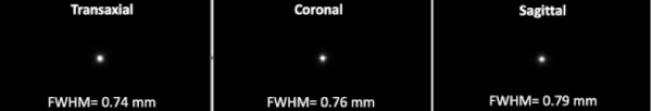
Objective 1: To develop a new continuous crystal PET system for simultaneous high spatial resolution (FWHM:~1mm) and sensitivity. System TOF resolution ~600ps for other organ background rejection.
Objective 2: To develop specific algorithms based on deep learning algorithms to be applied to data PET. This includes the software capable to deal with the location of gamma interaction events in the continuous scintillator crystal.
Objective 3: Optimize the continuous crystal PET system for breast cancer diagnostic and therapy follow-up.
Objective 4: Validation of the whole system performing a number of tests in accordance with the well-established NEMA standards that have been established for PET scanners
Objective 5: To develop a breast PET system compatible with high magnetic fields. In this sense, although we do not propose to test in this project the developed breast PET system within MRI devices, it is our intention to use fully compatible high magnetic field components that assures future development of PET-MRI multi-modal device.
DEEP-BREAST is a coordinated project between the Detectors for Molecular Imaging Laboratory group (DMIL) in charge for the hardware and Medical Imaging Reconstruction group (MIRG) in charge of the software aspects of the project. These research groups belong to the i3M.
PROJECT HARDWARE GOALS
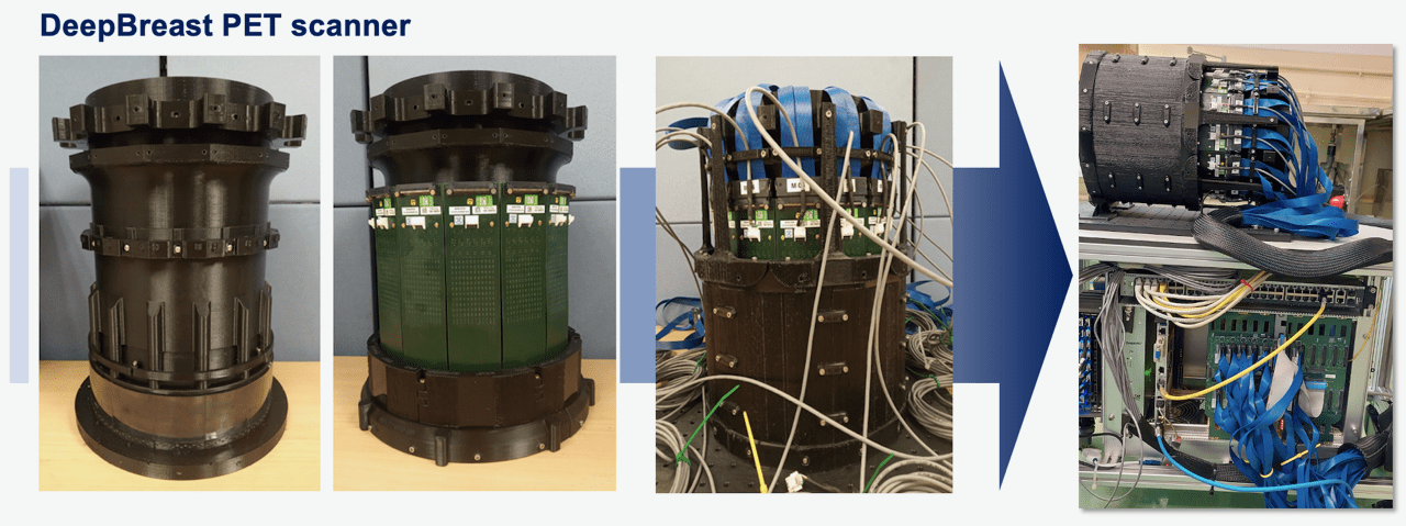
- To construct a breast PET system based on an edgeless design, including accurate determination of the gamma ray impact positions.
- To develop and integrate a readout electronics that minimizes capacitances of SiPM, together with ASICs devices.
- To develop all the mechanical support needed for PET detector integration
PROJECT SOFTWARE GOALS≤
- To develop a realistic digital breast phantom to be used for the generation of PET synthetic images for training the reconstruction algorithm.
- To develop the new software for image reconstruction, using deep learning techniques
Spanish Ministry of Science, Innovation and Universities: “Proyecto Nacional Retos i+d” DEEP-BREAST PID2019-107790RB-C21 and PID2019-107790RB-C22 funded by MCIN/ AEI /10.13039/501100011033