Research
Dental imaging with low-field magnetic resonance imaging (DentMRI)
* Funded by ATTRACT and European Commission under the H2020 program 2019
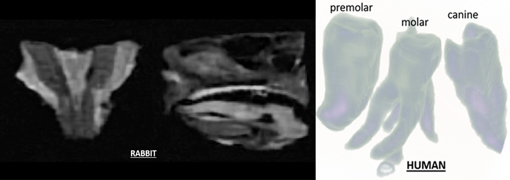
The main goal of the DentMRI project is to demonstrate a technology capable of simultaneous, high resolution imaging of soft and hard deep biological tissues, and which can be massively deployed in dental clinics within the next decade. Prior to the project start, we have been first to obtain high-resolution images of teeth (the hardest tissue present in the human body) in a Magnetic Resonance Imaging (MRI) scanner operating at low magnetic fields. Running at 0.3 T, well below the 1.5 T clinical standard, equipment costs are reduced by one to two orders of magnitude, a necessary condition for commercial viability.
In the scope of DentMRI, we will develop new electromagnetic pulse sequences for a test MRI scanner, as well as an image post-processing software application for diagnostic aid. We will use these to generate the first combined images of teeth and gum with diagnostic value, which will be assessed by professional odontologists. This test setup can accommodate objects of size up to 1 cubic centi-meter, so it is an additional goal of DentMRI to equip a human-head scanner with the radio-frequency electronics required for the pulse.