Research
Portable Photoacoustic Scanner: Towards a 3D virtual biopsy of melanoma.
Melanoma is one of the three most common malignant skin tumors, alongside basal cell and squamous cell carcinomas. Although less frequent, it accounts for nearly 90% of skin cancer-related deaths due to its high metastatic potential, driven by deep penetration of the primary tumor into blood and lymphatic vessels in the dermis. The Breslow thickness—the depth of tumor invasion—is the main prognostic indicator, currently measured ex vivo through biopsy and histopathology. Survival rates drop dramatically as Breslow thickness increases: from about 93% at 10 years for T1 tumors (<0.8 mm) to 50% for T4 tumors (>4 mm). This underscores the urgent need for new tools enabling early detection and routine clinical use.
This project proposes the development of the world’s first portable, low-cost 3D photoacoustic microscopy (PAM) scanner for early diagnosis and non-invasive monitoring of melanoma at the point of care.
Using near-infrared (NIR) laser excitation, PAM can reveal precise details of melanoma morphology and volumetric extension in vivo, at expected depths of 2 millimeters or more, offering clinicians valuable information for preliminary staging prior to biopsy—far beyond the superficial view of conventional dermoscopy. Additionally, a secondary visible laser can excite haemoglobin for angiographic imaging of neovascularization patterns. Recent results from a cost-effective, laser diode-based photoacoustic microscopy (PAM) system tested in vivo on mice have been published in the journal Ultrasonics in 2025.
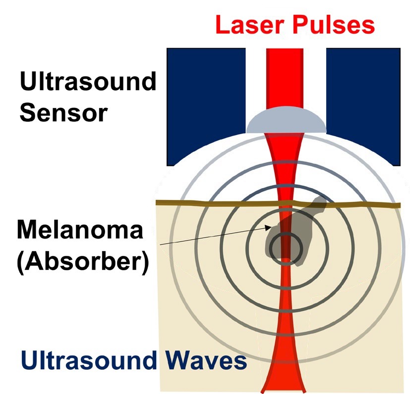
In general, Photoacoustic imaging (PAI) is an emerging modality that detects ultrasound waves generated inside tissue by laser absorption, combining the strengths of optical and ultrasound imaging. It enables high-contrast, volumetric 3D imaging at depths of several millimeters to centimeters, without ionizing radiation, pain, or invasive procedures. PAI visualizes endogenous chromophores such as melanin and haemoglobin, providing structural and functional information about tumors and vascular networks, and can be enhanced with exogenous contrast agents.
Project Objectives
- Design and build a portable, affordable clinical PAM prototype for melanoma diagnosis and monitoring at the point of care in close collaboration with the Instituto de Biomecánica de Valencia (IBV).
- Validate its utility and effectiveness in clinical trials with volunteer patients in collaboration with the Fundación Instituto Valenciano de Oncología (IVO) in collaboration with Dr. Onofre Sanmartín.
-
Assess potential medical benefits of early detection of melanoma, including minimization of surgical safety margins and improved detection of residual melanocytes post-excision and reduction of unnecessary biopsies through prior morphological and vascular characterization.
Beyond diagnosis, this technology holds promise for future therapeutic applications, such as photodynamic therapy and laser ablation guided by real-time 3D imaging, using the same scanning system with higher laser energy—though these applications fall outside the current project scope.
This breakthrough approach aims to democratize access to advanced melanoma imaging, enabling rapid, safe, and detailed molecular visualization for both clinical practice and biomedical research.
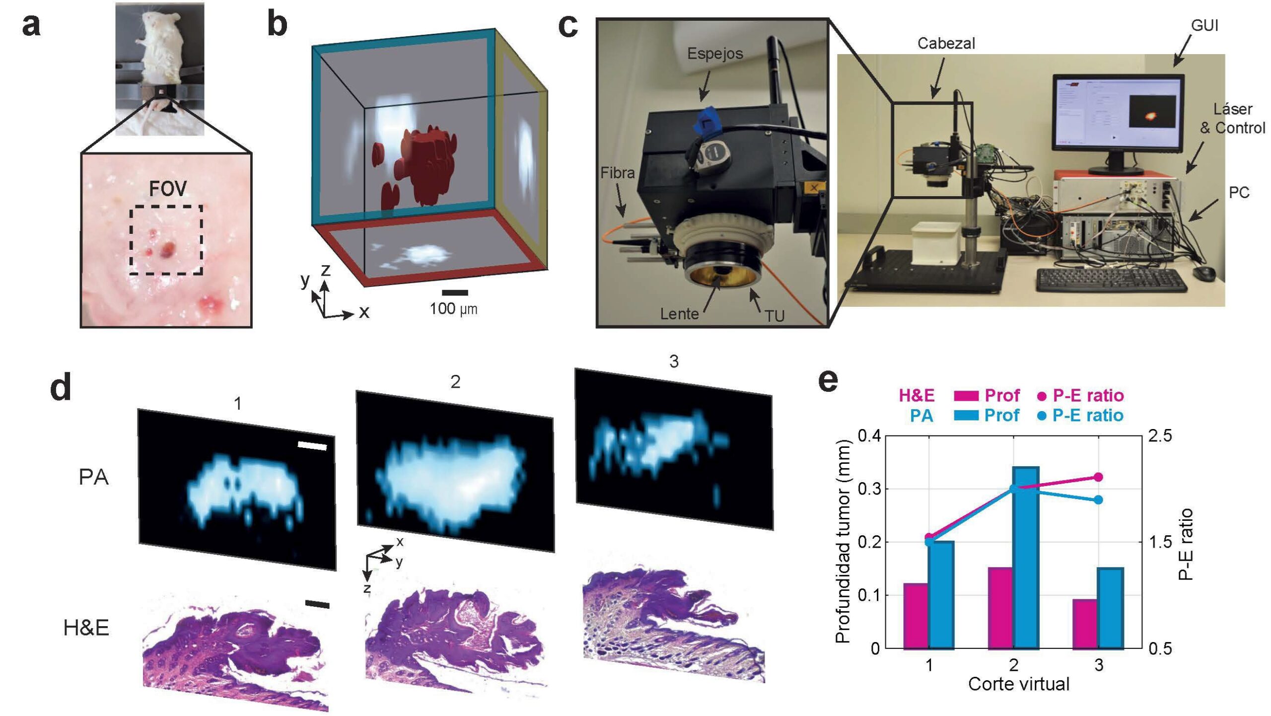
Results of preclinical 3D PAM scanner prototype in mice with melanocytic lesions [3].
Funding Acknowledgement
Project selected and action financed by the European Union through the European Regional Development Fund Program (ERDF) of the Valencian Region 2021-2027.
References:
1. Garbe et al. European consensus-based interdisciplinary guideline for melanoma. Part 1: Diagnostics: Update 2022, Eur J Cancer 170 (2022) 236–255.
https://doi.org/10.1016/j.ejca.2022.03.008
2. Lo et al., Long-Term Survival of Patients with Thin (T1) Cutaneous Melanomas: A Breslow Thickness Cut Point of 0.8 mm Separates Higher-Risk and Lower-Risk Tumors, Ann Surg Oncol 25 (2018) 894–902.
https://doi.org/10.1245/s10434-017-6325-1
3. Javier A. Navarro-Calvo, Alejandro Cebrecos, Adrián Arándiga, Laura Lorenzo-Rebenaque, Francisco Marco-Jiménez, José M. Benlloch, Francisco Camarena, Juan J. García-Garrigós, Cost-effective laser diode scanning 3D photoacoustic microscopy of melanocytic dermal tumors in situ, Ultrasonics, 158, 2026, 107831,
https://doi.org/10.1016/j.ultras.2025.107831
4. Lihong V. Wang, Song Hu, Photoacoustic Tomography: In Vivo Imaging from Organelles to Organs.Science335,1458-1462(2012). https://doi.org/10.1126/science.1216210